Back Rib Cage Muscles / Exercises to prevent back pain - CNN - Stretching out the muscles of the chest and the rib.. During breathing, these muscles normally tighten and pull the rib cage up. Located in upper mid stomach and also radiates to the back. Your chest expands and the lungs fill with air. It helps us to move as our muscles are attached to our bones. I no longer go to the emergency room (not recently.
These ribs are referred to as true ribs. I get muscle spasms in my stomach and rib cage muscles. Human rib cage anatomy model. The major abdominal muscles include the transverse abdominals ribs are attached to the breastbone that runs down the center of the chest. Stretching out the muscles of the chest and the rib.
Thoracic, Chest & Rib Pain | Aligned for Life from alignedforlife.co.uk Exhale and allow your rib cage and upper back come back to their natural position. The back muscles can be three types. If you were to develop well defined rib cage muscles, they would give off the appearance of fingers on your sides. The intercostal muscles are the muscles between the ribs. You'll need a bench and one lay back so your upper back is resting on the pad. These rib muscles automatically get worked when you do bench presses, push ups and dips, but a perform dumbbell pullovers to work the muscles along your rib cage. Did you know the rib cage plays a role in posture alignment? Things that need to addressed if hoping to make.
Human rib cage anatomy model.
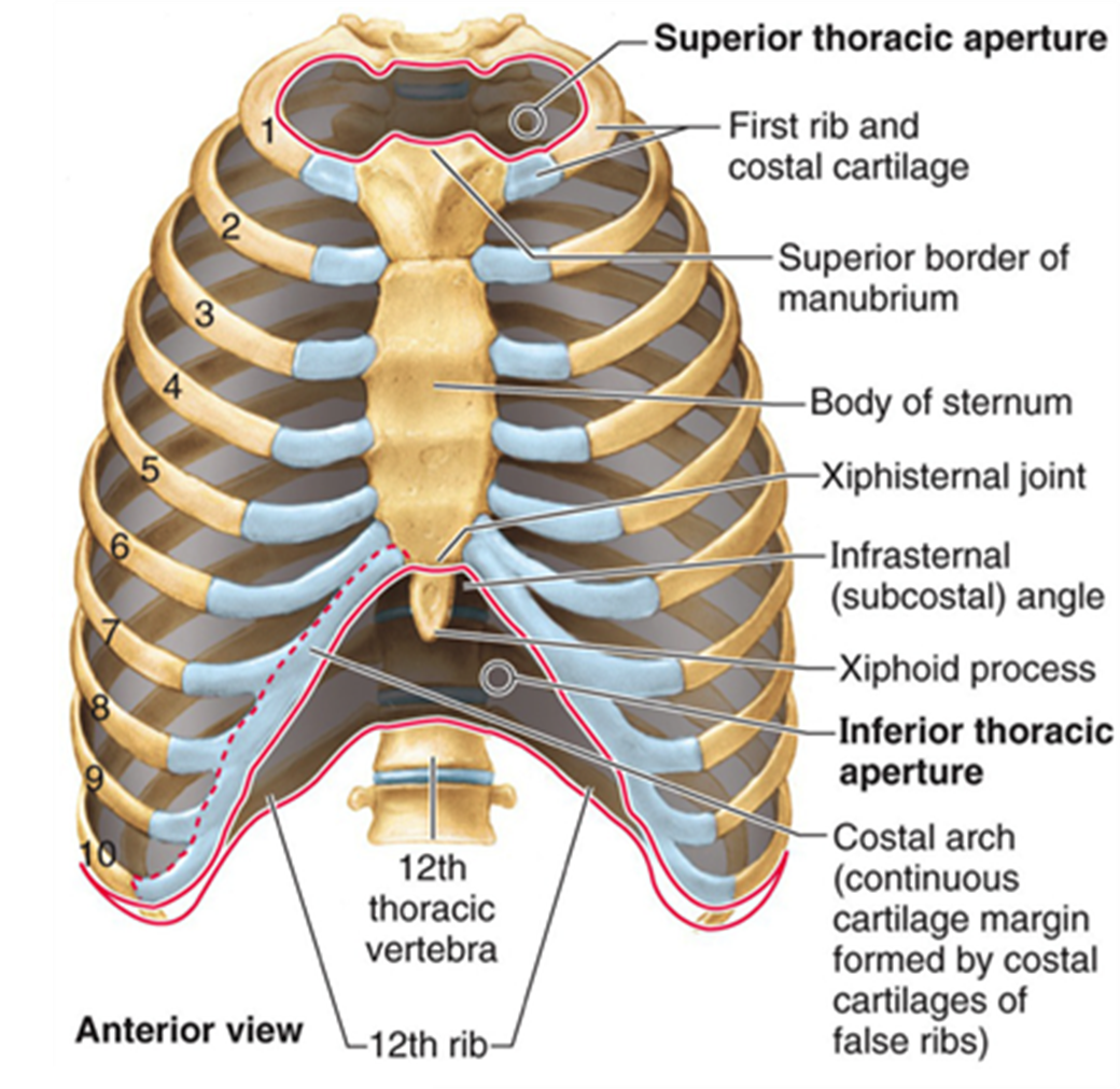
Injuries to your rib cage or muscles in your upper chest can cause rib pain ranging from a dull ache › get more: The back muscles can be three types. A typical human ribcage consists of 24 ribs, the sternum, costal cartilages, and the 12 thoracic vertebrae. Human male anatomy, 3/4 figure muscular and skeletal systems, back and front perspective views. Located in upper mid stomach and also radiates to the back. Learn more about the superficial muscles of the back with our learning materials that help you expand and test your knowledge in no time. External intercostals muscle are the outermost layer lies directly under the skin originate from the lower border. Spine, rib cage, back, torso. The superficial back muscles are the muscles found just under the skin. Consist of three layers of muscles external, internal, and innermost layer they combine to fill the space between the ribs. The muscles of the back that work together to support the spine, help keep the body upright and allow twist and bend in many directions. The rib cage has a major function in the respiratory system. How to stretch out the muscles of the chest & rib cage.
Learn about ribs muscle with free interactive flashcards. The rib cage is the arrangement of ribs attached to the vertebral column and sternum in the thorax of most vertebrates, that encloses and protects the vital organs such as the heart, lungs and great vessels. Try the stretches below to help improve your posture. Consist of three layers of muscles external, internal, and innermost layer they combine to fill the space between the ribs. Intermediate back muscles and c.
Thoracic Spine from www.spineuniverse.com The vertebral column, rib cage, and muscles of the back and abdomen. 3d rendering medical illustration of male interior brain anatomy. Intercostal muscles internal and external view. Stretching out the muscles of the chest and the rib. How to stretch out the muscles of the chest & rib cage. Within this group of back muscles you will find the latissimus dorsi, the trapezius, levator scapulae and the rhomboids. You may find that with practice, this natural, familiar, habitual position changes, and you acquire more distance between your ribs and pelvis. These ribs are referred to as true ribs.
Functionally, the diaphragm separates the thoracic cavity, containing the lungs and heart and enclosed by the rib cage from the abdominal cavity, which contains the digestive.
Human male anatomy, 3/4 figure muscular and skeletal systems, back and front perspective views. Also, many muscles between your ribs called intercostal muscles provide strength to your upper body and assist in breathing. The rib cage muscles consist of the obliques, intercostals and serratus anterior. Located in upper mid stomach and also radiates to the back. The cartilage is elastic and allows for expansion of the rib cage such as when taking a deep breath. Intercostal muscles internal and external view. How to stretch out the muscles of the chest & rib cage. At the front, they are attached by cartilage and at the back of the spine. Spine, rib cage, back, torso. The last time i had these was last friday night, and they lasted for two hours. The back muscles can be three types. Injuries to your rib cage or muscles in your upper chest can cause rib pain ranging from a dull ache › get more: Consist of three layers of muscles external, internal, and innermost layer they combine to fill the space between the ribs.
Consist of three layers of muscles external, internal, and innermost layer they combine to fill sudden severe pain, in the upper back or rib cage due to a direct blow or sudden impact to the chest or sudden increase in the physical activity. External intercostals muscle are the outermost layer lies directly under the skin originate from the lower border. This perfect spot lives in the thoracolumbar corner, a nook between your lowest rib and your spine — right where the stability of the rib cage and thoracic vertebrae gives. Muscle spasms felt within the rib cage may also be caused by the abdominal muscles. Stretching out the muscles of the chest and the rib.
Causes of Pain Under Right Rib Cage from i1.wp.com For the past three months, i've had a niggling pain/ache if you experience chest pain that is severe, that radiates to your arms, back or jaw, or is. The back muscles can be three types. Located in upper mid stomach and also radiates to the back. Back and rib muscles soreall education. The vertebral column, rib cage, and muscles of the back and abdomen. The muscles on your ribcage you are referring to are called the serratus anterior it is a muscle that originates on the surface of the 1st to 8th ribs at the side of the chest and inserts along the entire anterior length of the medial border of th. External intercostals muscle are the outermost layer lies directly under the skin originate from the lower border. Human rib cage anatomy model.
The back muscles can be three types.
Things that need to addressed if hoping to make. 3d rendering medical illustration of male interior brain anatomy. Stretching out the muscles of the chest and the rib. Injuries to your rib cage or muscles in your upper chest can cause rib pain ranging from a dull ache › get more: The muscles of the back that work together to support the spine, help keep the body upright and allow twist and bend in many directions. Human 3/4 body skeleton with muscles, veins and arteries. Clinically, chest muscle tightness generally follows a pattern of poor postures and upper back stiffness. Internal intercostal muscles sit directly underneath the external intercostals and help collapse the chest during breathing to exhale while the intercostal muscles do not connect directly to the spine, their stabilizing role in the rib cage assists in maintaining posture and keeping the back. The following general rules regarding actions can be. Clench your glutes to hold your torso parallel to. Suboccipital muscles (recti capitis posteriores major and minor, obliqui inferior and superior). Intercostal muscles are muscles that present within the rib cage. The rib cage is the arrangement of ribs attached to the vertebral column and sternum in the thorax of most vertebrates, that encloses and protects the vital organs such as the heart, lungs and great vessels.
Human 3/4 body skeleton with muscles, veins and arteries rib cage muscles. The muscles on your ribcage you are referring to are called the serratus anterior it is a muscle that originates on the surface of the 1st to 8th ribs at the side of the chest and inserts along the entire anterior length of the medial border of th.


Posting Komentar
0 Komentar